|
Share this:
Read this article in PDF format
Gallstone disease is a disease of hepato-biliary system, caused by cholesterol and/or bilirubin metabolic disorder, and characterized by formation of stones in the gallbladder and/or the biliary tract (1).
Gallstones are categorized as cholesterol, mixed, black pigment, or brown pigment stones (2). Cholesterol gallstones are the main type of gallstones and contain cholesterol as the major chemical constituent. Mixed cholesterol gallstones are composed of more than 50% cholesterol (2). Cholesterol and mixed gallstones are formed from biliary sludge, which stays for a long time in the gallbladder lumen. Biliary sludge consists of calcium bilirubinate granules, cholesterol monohydrate crystals, and biliary polymerized glycoprotein mucin (2-8). The dynamics of the transformation of biliary sludge into cholesterol stones has been shown as follows: diffused biliary sludge → surface biliary sludge → precipitating biliary sludge → a cholesterol gallstone without acoustic shadow → a cholesterol gallstone with acoustic shadow (9). The time of formation of cholesterol stones depends on the intensity of the precipitation processes of cholesterol monohydrate crystals in biliary sludge, and equals 3 to 36 months (2). Transformation proportion varies from 5 to 50% depending on the cause (2).
Black pigment stones are composed of either pure calcium bilirubinate or polymer-like complexes consisting of calcium, cooper, and large amounts of mucin glycoproteins.
Brown pigment stones are composed of calcium salts of unconjugated bilirubin, with varying amounts of cholesterol and protein. These stones are usually associated with infection (2).
The natural history of gallstones is typically defined in two separate groups of patients: those with symptomatic gallstones and those who are asymptomatic. The vast majorities of gallstones are asymptomatic and remain asymptomatic (2). As a rule, gallstone disease is asymptomatic, which is called “silent” stones. The rate of development of biliary pain is approximately 2% per year for 5 years and then decreases over time. The incidence of complications in patients with asymptomatic stones is low, and prophylactic removal of the gallbladder for this condition is not necessary (2). Patients who had an episode of uncomplicated biliary pain in the year, 38% per year had recurrent biliary pain (2). An incidence of recurrent biliary pain as high as 50% per year in those with symptomatic gallstones. 30% of patients with one episode of biliary pain will not have a recurrent episode (2). The estimated risk of developing biliary complications is estimated to be 1% to 2% per year and is thought to remain relatively constant over time (2).
If biliary pain occurs in the right upper abdomen and the gallbladder wall inflames, gallstone disease transforms into chronic calculous cholecystitis.
Chronic calculous cholecystitis is an inflammatory disease which affects the gallbladder wall and causes motoric-tonic dysfunctions of the biliary system, accompanied by presence of gallstones in the gallbladder lumen, and reveals as biliary pain (1, 3). The motoric dysfunction of the gallbladder can be caused by increased basal common bile duct resistance, muscle hypertrophy, and chronic inflammation in the gallbladder wall. Biliary colic is the most common presenting symptom of cholelithiasis. Approximately 75% of patients with symptomatic gallstone disease seek medical attention because of episodic abdominal pain (2). The syndrome of biliary colic is caused by intermittent obstruction of cystic duct by gallstones (2).
Cholecystectomy should be offered to patients only after significant biliary symptoms develop.
Diagnostic criteria of the chronic calculous cholecystitis
- Recurrent episodes of biliary pain in the right upper abdomen, sometimes in epigastrium, often with irradiation to the right scapular region. Biliary pains may be in the right hypochondrium, frequently or occasionally, of different intensity and duration, related to intake of fatty meals (1).
In addition, a biliary pain may occur with one or more of the following symptoms:
- regular or periodical feeling of bitter taste
- nausea, sometimes vomiting
- regular or periodical abdominal bloating and borborygmus
- unstable stool with constipation or diarrhea prevailing
- Impaired gallbladder emptying.
- According to ultrasound examination, thickening of the gallbladder wall up to 3-4 mm and presence of gallstones in the gallbladder lumen.
Causes of the gallbladder evacuation dysfunction, biliary pain and chronic inflammation in the gallbladder wall
- Pathology of the smooth muscle cells and epithelial cells in the gallbladder wall (high degree of COX-2 expression in the smooth muscle cells and epithelial cells of the gallbladder wall).
- Hypersecretion of glycoprotein biliary mucin into gallbladder lumen and increase of concentration of glycoprotein biliary mucin in the gallbladder bile over the point of polymerization (>2.0 mg/ml) (high degree of COX-2 expression in the epithelial cells of the gallbladder mucosa).
- Contractile dyscoordination of the gallbladder and cystic duct (high degree of COX-2 expression in the smooth muscle cells of the gallbladder and cystic duct).
- Increased basal cystic duct resistance (high degree of COX-2 expression in the smooth muscle cells of the cystic duct).
- Increased basal common bile duct resistance (high degree of COX-2 expression in the smooth muscle cells of the sphincter of Oddi).
Mechanism of development of pathologic disorders
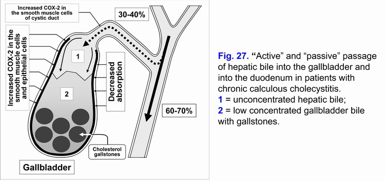
High degree of COX-2 expression in the smooth muscle cells of the gallbladder wall causes the decrease of the evacuation function of gallbladder and “active” passage of hepatic bile into the gallbladder (fig. 27).
Surplus COX-2 expression in the epithelial cells of the gallbladder mucosa makes for decrease of the absorption function of the gallbladder (decrease of water and biliary cholesterol absorption) and “passive” passage of the hepatic bile into the gallbladder (fig. 28). Also, gallstones volume in the gallbladder lumen may be the cause of decreased “active” and “passive” passage of the hepatic bile into the gallbladder (from 50% to 5%).
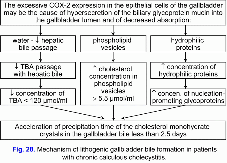
This is accompanied by decrease in concentration of total bile acids in the gallbladder bile and increase of concentration of biliary cholesterol in phospholipid vesicles, and causes disturbance in colloidal stability of gallbladder bile and precipitation of the cholesterol monohydrate crystals from unstable multilamellar aggregated phospholipid vesicles and calcium bilirubinate granules, i.e. formation of “lithogenic” gallbladder bile (fig. 28).
Also, increased COX-2 expression in the epithelial cells of the gallbladder mucosa activates the hypersecretion of glycoprotein mucin into the gallbladder lumen and gallbladder bile. The increase of the concentration of the glycoprotein biliary mucin in the gallbladder bile over 2.0 mg/ml causes its polymerization and formation of sites of the excessive viscosity and it is accompanied by rise of gallbladder bile viscosity. Precipitation of cholesterol monohydrate crystals and calcium bilirubinate granules in the sites of the excessive viscosity of polymerized glycoprotein biliary mucin causes the formation of biliary sludge, the increase in its echogenicity and its revelation during ultrasound examination.
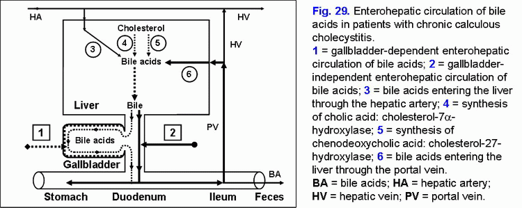
The decrease in “active” and “passive” passage of the hepatic bile into the gallbladder causes increase of passage of hepatic bile into duodenum and gallbladder-independent enterohepatic circulation of bile acids, biliary cholesterol and biliary bilirubin (fig. 29).
The increase in the gallbladder-independent enterohepatic circulation of bile acids causes increase of concentration of bile acids in the hepatocytes and the decrease in the accumulation function and excretion function of the liver (i.e. formation of chronic “bland” intrahepatic cholestasis) (fig. 29).
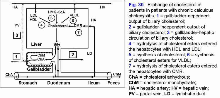
The increase of the gallbladder-independent enterohepatic circulation of biliary cholesterol causes increase of absorption of the biliary cholesterol in the small intestine, the biliary cholesterol entering hepatocytes and hypersecretion biliary cholesterol into hepatic bile (fig. 30).
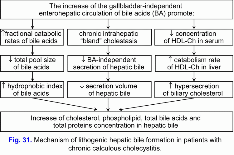
These two factors contribute to the formation of the “lithogenic” hepatic bile (fig. 31).
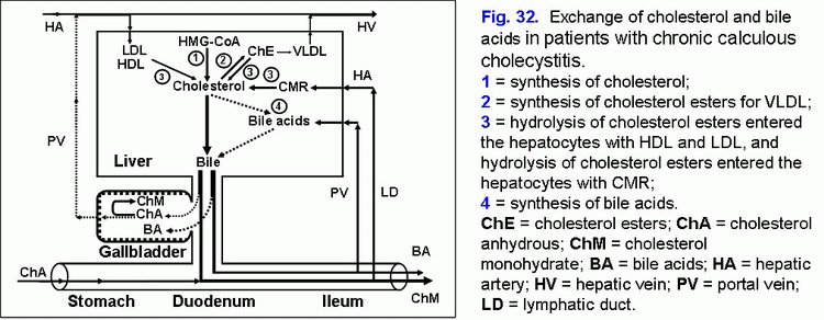
The decrease in the gallbladder-dependent output of biliary cholesterol and in the concentration of total bile acids in the gallbladder bile cause the formation of the “lithogenic” gallbladder bile and the precipitation of cholesterol monohydrate crystals in the gallbladder lumen among patients with chronic calculous cholecystitis (fig. 32).
Long-term storage of gallstones and chronic aseptic inflammation in the infundibulum mucosa of the gallbladder may cause the acute gallbladder obstruction of infundibulum and transformation of the chronic calculous cholecystitis into acute calculous cholecystitis, which need urgent surgical treatment.
Pathogenetic treatment of patients with chronic calculous cholecystitis
Accordingly, treatment of chronic calculous cholecystitis (with biliary pain) aiming for prophylactics of the acute calculous cholecystitis, duodeno-gastral reflux, antral atrophic (bile-acid-dependent) gastritis and chronic biliary pancreatitis includes:
- Celecoxib - 100 mg, 2 times a day after meal for 5-7 days, after which
- Ursodeoxycholic acid - 750 mg, once a day (in the evening) for 3 month.
Celecoxib is a selective inhibitor of COX-2. Inhibiting COX-2 activity in the smooth muscle cells of the gallbladder wall and cystic duct it brings relief of the biliary pain within 3-5 days, restoration of the evacuation function of the gallbladder and the gallbladder-dependent output of biliary cholesterol, “active” and “passive” passage of the hepatic bile into the gallbladder, and decrease in the gallbladder-independent enterohepatic circulation of bile acids, biliary cholesterol and biliary bilirubin.
Celecoxib, a selective inhibitor of COX-2, inhibiting COX-2 activity in the epithelial cells of the gallbladder mucosa causes inhibition of the glycoprotein mucin hypersecretion into the gallbladder lumen, concentration of glycoprotein biliary mucin in gallbladder bile and viscosity of gallbladder bile, which prevents formation of biliary sludge.
Low COX-2 activity in the epithelial cells of the gallbladder mucosa helps restoring the absorption function of the gallbladder (absorption of water and biliary cholesterol from phospholipid vesicles), which results in increase of concentration of total bile acids and decrease of concentration of biliary cholesterol in the gallbladder bile. Also, low COX-2 activity in the epithelial and smooth muscle cells of the gallbladder infundibulum helps lowering the risk of development of acute calculous cholecystitis.
Ursodeoxycholic acid, is a hydrophilic hepatoprotective bile acid. It helps in dissolving the cholesterol monohydrate crystals in the gallbladder, decrease of lithogenicity of gallbladder and hepatic bile, disappearance of the chronic “bland” intrahepatic cholestasis (i.e. results in the restoration of the accumulation and excretion functions of liver) and in some patients helps in dissolving cholesterol gallstones.
Celecoxib and Ursodeoxycholic acid, blocking main pathogenetic mechanisms of gallstones formation, help in slowing down the growth of cholesterol gallstones and lower the risk of acute calculous cholecystitis. In some patients the chronic calculous cholecystitis can transfer into the gallstone disease (without biliary pain) or the “silent” gallstones group.
Celecoxib is a selective inhibitor of COX-2. Inhibiting COX-2 activity in the smooth muscle cells of the biliary tract and the sphincter of Oddi it brings relief of the biliary pain within 3-5 days, restoration of the passage of the hepatic bile into the duodenum.
Celecoxib is a selective inhibitor of COX-2, inhibiting COX-2 activity in the epithelial cells of the biliary tract mucosa causes decrease in secretion of glycoprotein mucin into the biliary tract lumen, concentration of the glycoprotein biliary mucin in the hepatic bile and viscosity of hepatic bile, which prevents formation of biliary sludge and gallstones in the common hepatic duct and common bile duct. Low COX-2 activity in the epithelial cells and the smooth muscle cells of the biliary tract helps in lowering the risk of choledocholithiasis development.
Ursodeoxycholic acid (UDCA) is a hydrophilic hepatoprotective bile acid. It helps in dissolving the cholesterol monohydrate crystals in the biliary tract, decrease in lithogenicity of hepatic bile, disappearance of the chronic “bland” intrahepatic cholestasis (i.e. results in the restoration of the accumulation and excretion functions of liver), and in some patients helps in dissolving the biliary sludge in the biliary tract.
Ursodeoxycholic acid (UDCA) is a hydrophilic hepatoprotective bile acid, decreasing aggressive properties of bile, prevents development of chronic atrophic antral gastritis (duodenogastric reflux and bile reflux gastritis) and duodeno-gastroesophageal reflux (incompetence of Oddi's sphincter), chronic biliary pancreatitis (biliopancreatic reflux) or chronic spastic aseptic pancreatitis (pancreatic type III of sphincter of Oddi dysfunction).
Celecoxib and Ursodeoxycholic acid (UDCA), pathogenetically blocking main mechanisms of gallstone formation, help in prophylactics of gallstone formation in the biliary tract, and lower the risk of development of choledocholithiasis and chronic biliary pancreatitis (1-66).
Estimated effectiveness is 95%.
Remission period is 18-24 months.
Attention!!! Information for patients:
Before using this scheme of treatment please check the contraindications (below) and side effects of using pharmacological preparations of Celecoxib and Ursodeoxycholic acid, and obtain your doctor’s permission.
Contraindications for Celecoxib:
- allergic reactions (nettle-rash, bronchial spasm) to acetylsalicylic acid or other NSAIDs (in anamnesis);
- 3rd trimester of pregnancy;
- high sensitivity to sulphonamides;
- high sensitivity to any component of the preparation.
Contraindications for Ursodeoxycholic acid:
- high sensitivity to the preparation;
- acute inflammatory diseases of the gallbladder and the bile ducts;
- ulcerative colitis;
- Crone’s disease.
This web page does not bear any legal responsibility for the use of the proposed treatment schemes without consulting your doctor.
References:
- General practitioner: gastroenterology. 2002; 1: 22.
- Bilhartz L.E., Horton J.D. Gallstone disease and its complications. In: M. Feldman, B.F. Scharschmidt, M.H. Sleisenger, eds. Sleisenger and Fordtran’s Gastrointestinal and Liver Disease: Pathophysiology, Diagnosis, Management. 6th ed. Philadelphia: WB Saunders Company, 1998: 948-972.
- Ilchenko A.A., Delyukina O.V. Clinical role of biliary sludge. Consilium Medicum. Gastroenterology. 2005; 2.
- Lee S.P., Nicholls J.F. Nature and composition of biliary sludge. Gastroenterology. 1986; 90: 677-686.
- Carey M.C., Cahalane M.J. Whither biliary sludge? Gastroenterology. 1988; 95: 508-523.
- Wilkinson L.S., Levine T.S., Smith D., Chadwick S.J. Biliary sludge: can ultrasound reliably detect the presence of crystals in bile? Europ. J. Gastroenterol. Hepatol. 1996; 8: 999-1001.
- Jungst D., del Pozo R., Christoph S. et al. Quantification of biliary “sludge” in patients with cholesterol, mixed and pigment stones. Gastroenterology. 1994; 106(4): Abstr. 912.
- Jungst D., del Pozo R., Christoph S. et al. Sedimentation of biliary sludge. Effect on composition of gallbladder bile from patients with cholesterol, mixed and pigment stones. Scand. J. Gastroenterol. 1996; 31: 273-278.
- Inoue K., Fuchigami A., Higashide S. et al. Gallbladder sludge and stone formation in relation to contractile function after gastrectomy. Ann. Surg. 1992; 215: 19-26.
References (Celecoxib and UDCA):
- Chen XW, Cai JT. The impact of selective cycloxygenase-2 inhibitor celexibo on the formation of cholesterol gallstone. Zhonghua Nei Ke Za Zhi. 2003; 42(11): 797-9.
- Guarino MP, Carotti S, Sarzano M, Alloni R, Vanni M, Grosso M, Sironi G, Maffettone PL, Cicala M. Short-term ursodeoxycholic acid treatment improves gallbladder bile turnover in gallstone patients: a randomized trial. Neurogastroenterol Motil. 2005; 17(5): 680-6.
- Pazzi P, Petroni ML, Prandini N, Adam JA, Gullini S, Northfield TC, Jazrawi RP. Postprandial refilling and turnover: specific gallbladder motor function defects in patients with gallstone recurrence. Eur J Gastroenterol Hepatol. 2000; 12(7): 787-94.
- Ikegami T, Matsuzaki Y, Fukushima S, Shoda J, Olivier JL, Bouscarel B, Tanaka N. Suppressive effect of ursodeoxycholic acid on type IIA phospholipase A2 expression in HepG2 cells. Hepatology. 2005; 41(4): 896-905.
- Shoda J, Ueda T, Kawamoto T, Todoroki T, Asano T, Sugimoto Y, Ichikawa A, Maruyama T, Nimura Y, Tanaka N. Prostaglandin E receptors in bile ducts of hepatolithiasis patients and the pathobiological significance for cholangitis. Clin Gastroenterol Hepatol. 2003; 1(4): 285-96.
- Shoda J, Kano M, Asano T, Irimura T, Ueda T, Iwasaki R, Furukawa M, Kamiya J, Nimura Y, Todoroki T, Matsuzaki Y, Tanaka N. Secretory low-molecular-weight phospholipases A2 and their specific receptor in bile ducts of patients with intrahepatic calculi: factors of chronic proliferative cholangitis. Hepatology. 1999; 29(4): 1026-36.
- Tomida S, Abei M, Yamaguchi T, Matsuzaki Y, Shoda J, Tanaka N, Osuga T. Long-term ursodeoxycholic acid therapy is associated with reduced risk of biliary pain and acute cholecystitis in patients with gallbladder stones: a cohort analysis. Hepatology. 1999; 30(1): 6-13.
- Kano M, Shoda J, Irimura T, Ueda T, Iwasaki R, Urasaki T, Kawauchi Y, Asano T, Matsuzaki Y, Tanaka N. Effects of long-term ursodeoxycholate administration on expression levels of secretory low-molecular-weight phospholipases A2 and mucin genes in gallbladders and biliary composition in patients with multiple cholesterol stones. Hepatology. 1998; 28(2): 302-13.
- Shoda J, Ueda T, Ikegami T, Matsuzaki Y, Satoh S, Kano M, Matsuura K, Tanaka N. Increased biliary group II phospholipase A2 and altered gallbladder bile in patients with multiple cholesterol stones. Gastroenterology. 1997; 112(6): 2036-47.
- Carotti S, Guarino MP, Cicala M, Perrone G, Alloni R, Segreto F, Rabitti C, Morini S. Effect of ursodeoxycholic acid on inflammatory infiltrate in gallbladder muscle of cholesterol gallstone patients. Neurogastroenterol Motil. 2010.
- Guarino MP, Carotti S, Morini S, Perrone G, Behar J, Altomare A, Alloni R, Caviglia R, Emerenziani S, Rabitti C, Cicala M. Decreased number of activated macrophages in gallbladder muscle layer of cholesterol gallstone patients following ursodeoxycholic acid. Gut. 2008; 57(12): 1740-1.
- Jüngst C, Sreejayan N, Zündt B, Müller I, Spelsberg FW, Hüttl TP, Kullak-Ublick GA, del Pozo R, Jüngst D, von Ritter C. Ursodeoxycholic acid reduces lipid peroxidation and mucin secretagogue activity in gallbladder bile of patients with cholesterol gallstones. Eur J Clin Invest. 2008; 38(9): 634-9.
- Spier BJ, Pfau PR, Lorenze KR, Knechtle SJ, Said A. Risk factors and outcomes in post-liver transplantation bile duct stones and casts: A case-control study. Liver Transpl. 2008; 14(10): 1461-5.
- Guarino MP, Cong P, Cicala M, Alloni R, Carotti S, Behar J. Ursodeoxycholic acid improves muscle contractility and inflammation in symptomatic gallbladders with cholesterol gallstones. Gut. 2007; 56(6): 815-20.
- Mas MR, Comert B, Mas N, Yamanel L, Ozotuk H, Tasci I, Jazrawi RP. Effects of long term hydrophilic bile acid therapy on in vitro contraction of gallbladder muscle strips in patients with cholesterol gallstones. World J Gastroenterol. 2007; 13(32): 4336-9.
- Jüngst C, Sreejayan N, Eder MI, von Stillfried N, Zündt B, Spelsberg FW, Kullak-Ublick GA, Jüngst D, von Ritter C. Lipid peroxidation and mucin secretagogue activity in bile of gallstone patients. Eur J Clin Invest. 2007; 37(9): 731-6.
- Itoh S, Kono M, Akimoto T. Psoriasis treated with ursodeoxycholic acid: three case reports. Clin Exp Dermatol. 2007; 32(4): 398-400.
- Beuers U. Drug insight: Mechanisms and sites of action of ursodeoxycholic acid in cholestasis. Nat Clin Pract Gastroenterol Hepatol. 2006; 3(6): 318-28.
- Colecchia A, Mazzella G, Sandri L, Azzaroli F, Magliuolo M, Simoni P, Bacchi-Reggiani ML, Roda E, Festi D. Ursodeoxycholic acid improves gastrointestinal motility defects in gallstone patients. World J Gastroenterol. 2006; 12(33): 5336-43.
- Pemberton PW, Aboutwerat A, Smith A, Warnes TW. Ursodeoxycholic acid in primary biliary cirrhosis improves glutathione status but fails to reduce lipid peroxidation. Redox Rep. 2006; 11(3): 117-23.
- Jeong HJ, Kim CG. Pretreatment with ursodeoxycholic acid (UDCA) as a novel pharmacological intervention in hepatobiliary scintigraphy. Yonsei Med J. 2005; 46(3): 394-8.
- Fischer S, Müller I, Zündt BZ, Jüngst C, Meyer G, Jüngst D. Ursodeoxycholic acid decreases viscosity and sedimentable fractions of gallbladder bile in patients with cholesterol gallstones. Eur J Gastroenterol Hepatol. 2004; 16(3): 305-11.
- Sauter GH, Thiessen K, Parhofer KG, Jüngst C, Fischer S, Jüngst D. Effects of ursodeoxycholic acid on synthesis of cholesterol and bile acids in healthy subjects. Digestion. 2004; 70(2): 79-83.
- Xiao ZL, Biancani P, Carey MC, Behar J. Hydrophilic but not hydrophobic bile acids prevent gallbladder muscle dysfunction in acute cholecystitis. Hepatology. 2003; 37(6): 1442-50.
- Tazuma S, Nishioka T, Ochi H, Hyogo H, Sunami Y, Nakai K, Tsuboi K, Asamoto Y, Sakomoto M, Numata Y, Kanno K, Yamaguchi A, Kobuke T, Komichi D, Nonaka Y, Chayama K. Impaired gallbladder mucosal function in aged gallstone patients suppresses gallstone recurrence after successful extracorporeal shockwave lithotripsy. J Gastroenterol Hepatol. 2003; 18(2): 157-61.
- Gunsar C, Melek M, Karaca I, Sencan A, Mir E, Ortac R, Canan O. The biochemical and histopathological effects of ursodeoxycholic acid and metronidazole on total parenteral nutrition-associated hepatic dysfunction: an experimental study. Hepatogastroenterology. 2002; 49(44): 497-500.
- Xiao ZL, Rho AK, Biancani P, Behar J. Effects of bile acids on the muscle functions of guinea pig gallbladder. Am J Physiol Gastrointest Liver Physiol. 2002; 283(1): G87-94.
- Kano M, Shoda J, Satoh S, Kobayashi M, Matsuzaki Y, Abei M, Tanaka N. Increased expression of gallbladder cholecystokinin: a receptor in prairie dogs fed a high-cholesterol diet and its dissociation with decreased contractility in response to cholecystokinin. J Lab Clin Med. 2002; 139(5): 285-94.
- Wang DQ, Tazuma S. Effect of beta-muricholic acid on the prevention and dissolution of cholesterol gallstones in C57L/J mice. J Lipid Res. 2002; 43(11): 1960-8.
- Lukivskaya OY, Maskevich AA, Buko VU. Effect of ursodeoxycholic acid on prostaglandin metabolism and microsomal membranes in alcoholic fatty liver. Alcohol. 2001; 25(2): 99-105.
- Bomzon A, Ljubuncic P. Ursodeoxycholic acid and in vitro vasoactivity of hydrophobic bile acids. Dig Dis Sci. 2001; 46(9): 2017-24.
- Sunami Y, Tazuma S, Kajiyama G. Gallbladder dysfunction enhances physical density but not biochemical metastability of biliary vesicles. Dig Dis Sci. 2000; 45(12): 2382-91.
- Ljubuncic P, Said O, Ehrlich Y, Meddings JB, Shaffer EA, Bomzon A. On the in vitro vasoactivity of bile acids. Br J Pharmacol. 2000; 131(3): 387-98.
- Nishioka T, Tazuma S, Yamashita G, Kajiyama G. Partial replacement of bile salts causes marked changes of cholesterol crystallization in supersaturated model bile systems. Biochem J. 1999; 340 ( Pt 2): 445-51.
- Sinisalo J, Vanhanen H, Pajunen P, Vapaatalo H, Nieminen MS. Ursodeoxycholic acid and endothelial-dependent, nitric oxide-independent vasodilatation of forearm resistance arteries in patients with coronary heart disease. Br J Clin Pharmacol. 1999; 47(6): 661-5.
- van de Heijning BJ, van de Meeberg PC, Portincasa P, Doornewaard H, Hoebers FJ, van Erpecum KJ, Vanberge-Henegouwen GP. Effects of ursodeoxycholic acid therapy on in vitro gallbladder contractility in patients with cholesterol gallstones. Dig Dis Sci. 1999; 44(1): 190-6.
- Mendez-Sanchez N, Brink MA, Paigen B, Carey MC. Ursodeoxycholic acid and cholesterol induce enterohepatic cycling of bilirubin in rodents. Gastroenterology. 1998; 115(3): 722-32.
- Benedetti A, Alvaro D, Bassotti C, Gigliozzi A, Ferretti G, La Rosa T, Di Sario A, Baiocchi L, Jezequel AM. Cytotoxicity of bile salts against biliary epithelium: a study in isolated bile ductule fragments and isolated perfused rat liver. Hepatology. 1997; 26(1): 9-21.
- Ohtake M, Sandoh N, Sakaguchi T, Tsukada K, Hatakeyama K. Enhancement of portal blood flow by ursodesoxycholic acid in partially hepatectomized rats. Surg Today. 1996; 26(2): 142-4.
- Fahey DA, Carey MC, Donovan JM. Bile acid/phosphatidylcholine interactions in mixed monomolecular layers: differences in condensation effects but not interfacial orientation between hydrophobic and hydrophilic bile acid species. Biochemistry. 1995; 34(34): 10886-97.
- Bouscarel B, Ceryak S, Robins SJ, Fromm H. Studies on the mechanism of the ursodeoxycholic acid-induced increase in hepatic low-density lipoprotein binding. Lipids. 1995; 30(7): 607-17.
- Bomzon A, Ljubuncic P. Bile acids as endogenous vasodilators? Biochem Pharmacol. 1995; 49(5): 581-9.
- Jazrawi RP, Pazzi P, Petroni ML, Prandini N, Paul C, Adam JA, Gullini S, Northfield TC. Postprandial gallbladder motor function: refilling and turnover of bile in health and in cholelithiasis. Gastroenterology. 1995; 109(2): 582-91.
- Pak JM, Adeagbo AS, Triggle CR, Shaffer EA, Lee SS. Mechanism of bile salt vasoactivity: dependence on calcium channels in vascular smooth muscle. Br J Pharmacol. 1994; 112(4): 1209-15.
- Sasaki H, Tazuma S, Kajiyama G. Effects of 16,16-dimethyl prostaglandin E2 on biliary mucous glycoprotein and gallstone formation in guinea pigs. Scand J Gastroenterol. 1993; 28(6): 495-9.
- Mizuno S, Tazuma S, Kajiyama G. Stabilization of biliary lipid particles by ursodeoxycholic acid. Prolonged nucleation time in human gallbladder bile. Dig Dis Sci. 1993; 38(4): 684-93.
- Pak JM, Lee SS. Vasoactive effects of bile salts in cirrhotic rats: in vivo and in vitro studies. Hepatology. 1993; 18(5): 1175-81.
- Das JB, Cosentino CM, Levy MF, Ansari GG, Raffensperger JG. Early hepatobiliary dysfunction during total parenteral nutrition: an experimental study. J Pediatr Surg. 1993; 28(1): 14-8.
- Fromm H, Malavolti M. Bile acid dissolution therapy of gallbladder stones. Baillieres Clin Gastroenterol. 1992; 6(4): 689-95.
- Tazuma S, Sasaki H, Mizuno S, Sagawa H, Hashiba S, Horiuchi I, Kajiyama G. Effect of ursodeoxycholic acid administration on nucleation time in human gallbladder bile. Gastroenterology. 1989; 97(1): 173-8.
|